Reading an Mri Brain Scan Is Subjective
| Magnetic Resonance Imaging (MRI) of the Brain and Spine: Basics | ||||||||||||||||||||||||
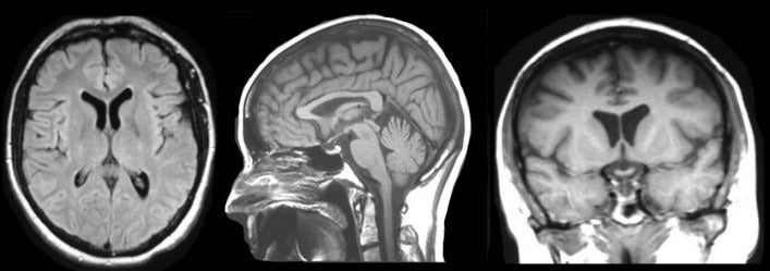
| Magnetic resonance imaging (MRI) is 1 of the most commonly used tests in neurology and neurosurgery. MRI provides exquisite detail of brain, spinal cord and vascular anatomy, and has the advantage of beingness able to visualize anatomy in all three planes: axial, sagittal and coronal (see the case image below). | ||||||||||||||||||||||||
| | ||||||||||||||||||||||||
| MRI has an reward over CT in existence able to find flowing blood and ambiguous vascular malformations. It tin can also detect demyelinating disease, and has no beam-hardening artifacts such as can exist seen with CT. Thus, the posterior fossa is more easily visualized on MRI than CT. Imaging is too performed without any ionizing radiation. | ||||||||||||||||||||||||
| PHYSICS OF MRI MRI is based on the magnetization properties of diminutive nuclei. A powerful, compatible, external magnetic field is employed to marshal the protons that are usually randomly oriented within the water nuclei of the tissue existence examined. This alignment (or magnetization) is next perturbed or disrupted past introduction of an external Radio Frequency (RF) free energy. The nuclei return to their resting alignment through various relaxation processes and in and so doing emit RF free energy. After a sure menses post-obit the initial RF, the emitted signals are measured. Fourier transformation is used to convert the frequency data independent in the bespeak from each location in the imaged plane to corresponding intensity levels, which are then displayed as shades of gray in a matrix arrangement of pixels. By varying the sequence of RF pulses applied & collected, different types of images are created. Repetition Time (TR) is the amount of time between successive pulse sequences applied to the same slice. Time to Repeat (TE) is the time betwixt the delivery of the RF pulse and the receipt of the echo signal. Tissue tin can be characterized by two different relaxation times � T1 and T2. T1 (longitudinal relaxation fourth dimension) is the time abiding which determines the rate at which excited protons render to equilibrium. It is a measure of the time taken for spinning protons to realign with the external magnetic field. T2 (transverse relaxation time) is the time constant which determines the rate at which excited protons reach equilibrium or become out of stage with each other. It is a measure of the time taken for spinning protons to lose phase coherence amid the nuclei spinning perpendicular to the main field. | ||||||||||||||||||||||||
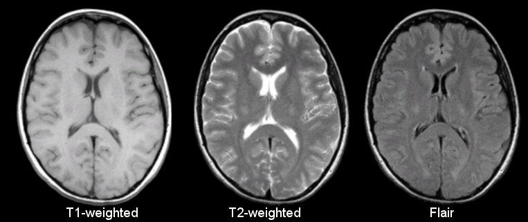
| MRI IMAGING SEQUENCES The virtually mutual MRI sequences are T1-weighted and T2-weighted scans. T1-weighted images are produced by using curt TE and TR times. The contrast and brightness of the image are predominately determined by T1 properties of tissue. Conversely, T2-weighted images are produced by using longer TE and TR times. In these images, the dissimilarity and brightness are predominately determined by the T2 properties of tissue. In general, T1- and T2-weighted images can be easily differentiated by looking the CSF. CSF is dark on T1-weighted imaging and vivid on T2-weighted imaging. A third commonly used sequence is the Fluid Attenuated Inversion Recovery (Flair) . The Flair sequence is like to a T2-weighted image except that the TE and TR times are very long. Past doing then, abnormalities remain bright but normal CSF fluid is attenuated and made night. This sequence is very sensitive to pathology and makes the differentiation between CSF and an abnormality much easier. | ||||||||||||||||||||||||
Above : Most common MRI Sequences and their Guess TR and TE times. | ||||||||||||||||||||||||
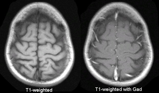
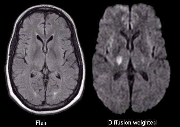
| T1-weighted imaging can also be performed while infusing Gadolinium (Gad) . Gad is a not-toxic paramagnetic contrast enhancement amanuensis. When injected during the scan, Gad changes betoken intensities by shortening T1. Thus, Gad is very brilliant on T1-weighted images. Gad enhanced images are especially useful in looking at vascular structures and breakdown in the blood-brain barrier [eastward.g., tumors, abscesses, inflammation (herpes simplex encephalitis, multiple sclerosis, etc.)]. Diffusion weighted imaging (DWI) is designed to find the random movements of water protons. Water molecules lengthened relatively freely in the extracellular space; their motion is significantly restricted in the intracellular space. Spontaneous movements, referred to every bit diffusion, chop-chop become restricted in ischemic brain tissue. During ischemia, the sodium - potassium pump shuts downward and sodium accumulates intracellularly. Water then shifts from the extracellular to the intracellular space due to the osmotic gradient. As h2o motility becomes restricted intracellularly, this results in an extremely bright bespeak on DWI. Thus, DWI is an extremely sensitive method for detecting acute stroke. | ||||||||||||||||||||||||
| Comparing of T1 vs. T2 vs. Flair (Encephalon) | ||||||||||||||||||||||||
| ||||||||||||||||||||||||
| Comparison of T1 vs. T1 with Gadolinium
| ||||||||||||||||||||||||
| | ||||||||||||||||||||||||
| Comparison of Flair vs. Improvidence-weighted | ||||||||||||||||||||||||
| | ||||||||||||||||||||||||
| | ||||||||||||||||||||||||
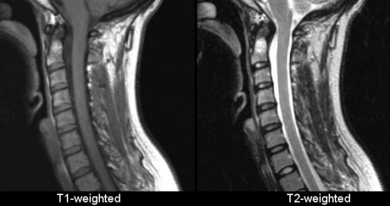
| Comparing of T1 vs. T2 - Spine | ||||||||||||||||||||||||
| NEUROLOGICAL INDICATIONS FOR CRANIAL MRI ● Vascular (ischemic and hemorrhagic stroke, AVM, aneurysm, venous thrombosis) ● Tumor (chief CNS and metastatic) ● Infection (abscess, cerebritis, encephalitis, meningitis) ● Inflammatory/Demyelinating Lesions (multiple sclerosis, sarcoidosis, etc.) ● Trauma (epidural hematoma, subdural hematoma, contusion) ● Hydrocephalus ● Congenital Malformations | ||||||||||||||||||||||||
| LIMITATIONS OF MRI ● Subject to motion artifact ● Junior to CT in detecting acute hemorrhage ● Inferior to CT in detection of bony injury ● Requires prolonged acquisition time for many images | ||||||||||||||||||||||||
| CONTRAINDICATIONS TO MRI In that location are few contraindications to MRI. Nearly contraindications to MRI tin be divided into the following groups: ● Implanted devices and other metal devices - Pacemakers and other implanted electronic devices ● Intraocular metal foreign bodies - Screening CT of the orbits if history suggests possible metallic foreign trunk in the heart ● Unstable patients (almost resuscitation equipment cannot be brought into the scanning room) ● Pregnancy (relative contraindication due to unknown furnishings on the fetus) ● Other � severe agitation, or claustrophobia (may require anesthesia assistance) |
Source: https://case.edu/med/neurology/NR/MRI%20Basics.htm
0 Response to "Reading an Mri Brain Scan Is Subjective"
Post a Comment